
Sub-Micron 3D X-ray Microscopy
ZEISS Xradia Versa 610
3D X-ray Microscopy for Faster Sub-Micron Imaging of Intact Samples
Sub-micron X-ray microscopy (XRM) offers special features that make this technique a powerful tool for industrial and academical research. Lab-based XRM Xradia Versa 610 delivers high resolution and contrast in three dimensions, in a non-destructive way, thus pushing the limit well beyond that of classical X-ray tomography or micro-CT. Moreover, it is possible to describe structural analysis and follow microstructural changes over time, over a wide range of length scales through in situ and 4D imaging. With this strength, it is possible to characterize an ever-increasing range of materials, from biological samples to batteries, semiconductors, rocks, composite materials, even performing dynamic experiments.
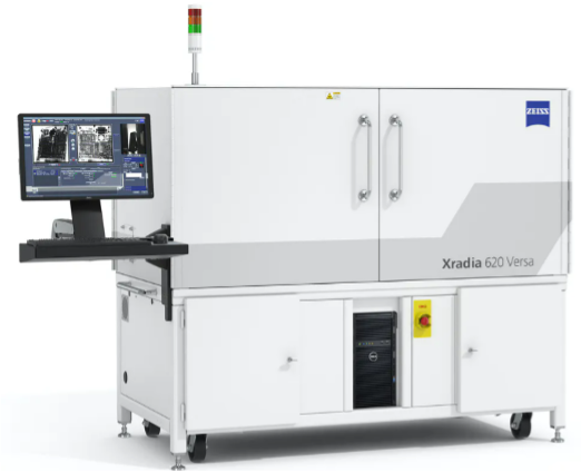
Xradia Versa 610 (https://www.zeiss.com/microscopy/int/products/x-ray-microscopy/zeiss-xradia-610-and-620-versa.html)
The new concept of Resolution at a Distance (Raad)
The Highest Resolution at large distances
ZEISS Xradia Versa 610 provides a dual-stage magnification, by adding an optical magnification step to the geometrical one, extending in this way the resolution limit also for big object. This configuration, illustrated in the following Figure, takes the name of Resolution at a Distance (RaaD).
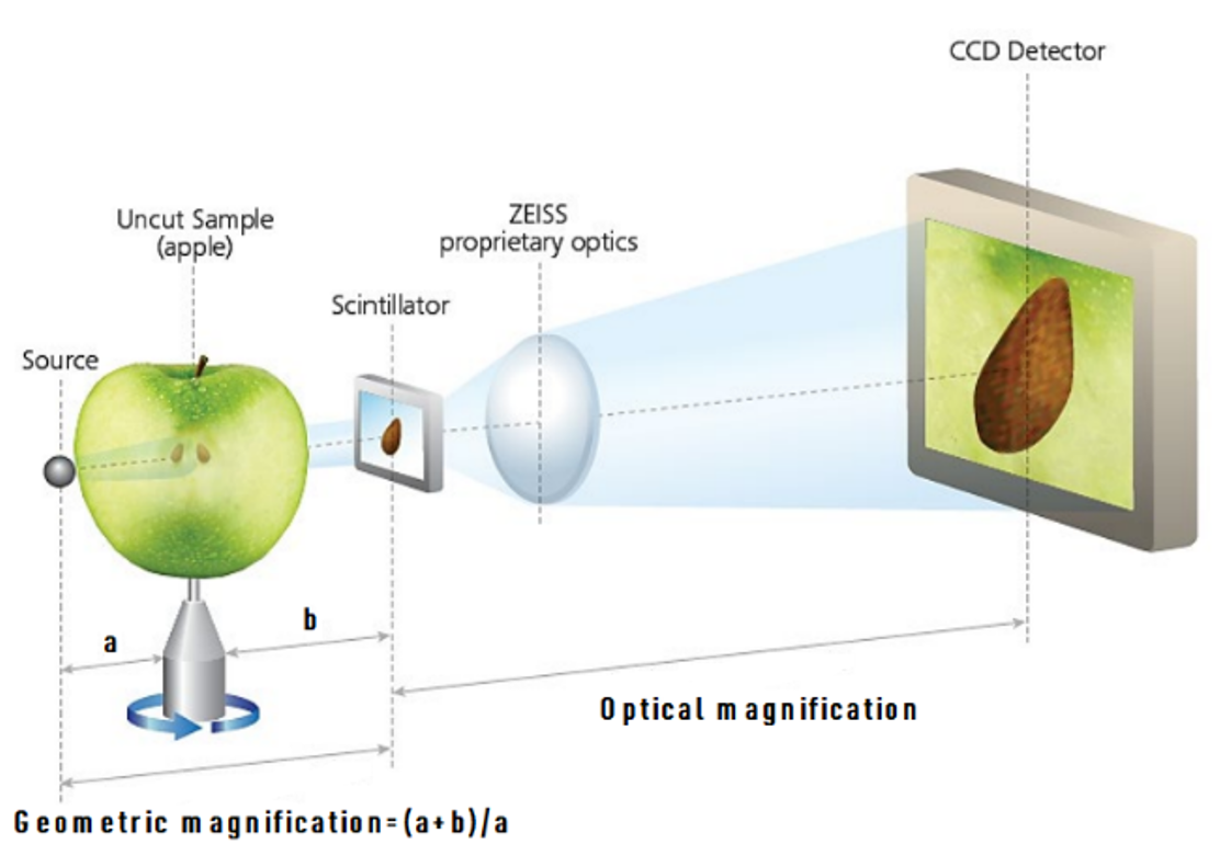
Schematic representation of the ZEISS Resolution at a Distance Sub-micron XRM architecture.
(Source: https://www.zeiss.com/microscopy/int/products/x-ray-microscopy/zeiss-xradia-610-and-620-versa.html.)
In this configuration the projections are at first enlarged through geometric magnification, as in conventional microCT, the magnified X-rays impinge on the scintillator, that is placed on the top of the objective, and are transformed into visible light, that is subsequently magnified by an optical lens system. With more X-ray photons available, the ZEISS Xradia 600 Series Versa provides faster time to results for the widest range of sample sizes and types, without compromising resolution.
s shown in the next Figure, the importance of the sub-micron XRM comes from the possibility to obtain a non-destructive 3D imaging within a large resolution range while, at the same time, requiring a limited sample preparation, thus being able to close the gap with more complex and resolute techniques, such as FIB-SEM or TEM tomography.
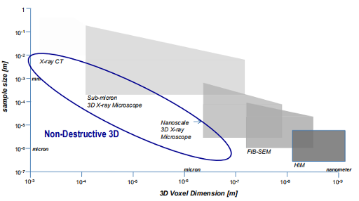
3D Imaging technologies range ranges. (Source: F.-X. Masson, G. Beaudoin and D. Laurendeau, "Quantification of the morphology of gold grains in 3D using X-ray microscopy and SEM photogrammetry," Journal of Sedimentary Research, vol. 3, no. 90, p. 286–296, 2020).
Xradia Versa 610 Highlights
Moving beyond the limits of Traditional Micro-CT
- Non-destructive sub-micron scale microscopy of intact sample
- 3D representation over a range of scales and samples
- Resolution-at-a-Distance (RaaD) adding an optical magnification step to the geometrical one, extending the resolution limit also for big object
- High resolution across a broad range of sample types, sizes, and working distances
- True spatial resolution of 500 nm with a minimum achievable voxel size of 40 nm
- In situ imaging for non-destructive 4D analysis in controlled environments